Medical Imaging
Our in-house Consultant Radiologists, Medical Sonologists, Medical Radiographers, Cardiologists are experts in medical imaging/radiology. They use their expertise coupled with cutting-edge technologies to produce accurate and more realistic images for medical diagnosis.
- Ultrasonography
This diagnostic medical imaging technique uses high-frequency sound waves to create an image of part of the body on the screen. It does not use ionising radiation. It is safe for everyone including, pregnant women, Children, Adults- Male and Female- and the Elderly. Some of our Ultrasound Services include Pelvic scan, Abdominal scan, Pregnancy scan, Children scan, Breast scan, Transvaginal scan, Thyroid scan, Prostate scan, Eye (Ocular) scan, Vascular (Obstetrics and other parts Doppler) scan, and Heart scan (Echocardiography).
- High-quality image resolution.
- More realistic 5D images.
- More realistic 5D images. Accurate foetal weight estimation
- Extensive foetal central nervous system assessment using 5D CNS+ support.
- E-Strain Elastoscan; for accurate diagnosis of breast, Liver and thyroid masses.
- Adjustable built-in integrated ultrasound examination couch; for maximum patient comfort during scans.
- Full range of transducers (Convex, Volume, Linear, Transrectal, Transvaginal, and Cardiac probes) configured with latest technologies.
-Echocardiography
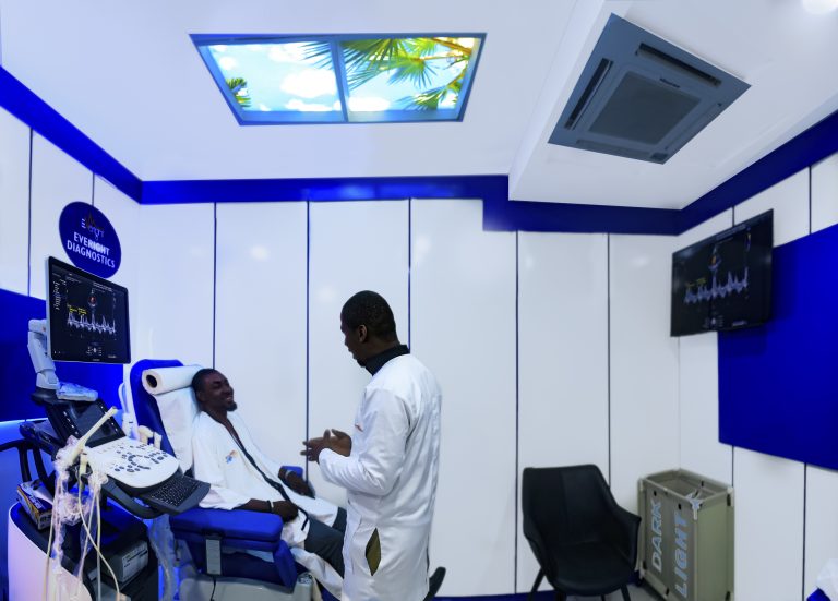
This is an ultrasound scan of the heart. It is used to check anomalies (congenital and acquired) concerning the chambers, the valves, the wall and the enclosing sac (pericardium) of the heart. It is safe for everyone (children and adults).
ADULT ECHOCARDIOGRAPHY.
Adult echocardiography is ultrasonography used to evaluate the anatomical structure and function of the heart and great vessels in Adults. An echocardiogram is a test that uses sound waves (ultrasound) to create images of the heart. At Everight, we have a dedicated 4D Ultrasound Machine with experienced Consultant Cardiologists for Echocardiography examinations. Some indications/reasons for doing an Echocardiography will include the following:
- Chest pain, shortness of breath, syncope, palpitations, transient ischemic attack, stroke, and peripheral embolic event
- Previous diagnostic tests such as cardiac enzymes, electrocardiogram, and chest X-ray indicating cardiac disease
- Premature ventricular contraction
- Arrhythmias
- Suspected pulmonary artery hypertension
- To guide management in patients diagnosed with pulmonary artery hypertension
- Patient in shock with an uncertain or suspected cardiac cause
- For diagnosis of valvular heart disease
- To guide management in patients diagnosed with valvular heart disease
- Patients with suspicion of hypertensive heart disease, prosthetic valve dysfunction, infective endocarditis, heart failure, congenital heart disease, cardiac mass, pericardial disease, aortic aneurysm, aortic dissection, or cardiovascular source of embolus
PAEDIATRIC ECHOCARDIOGRAPHY.
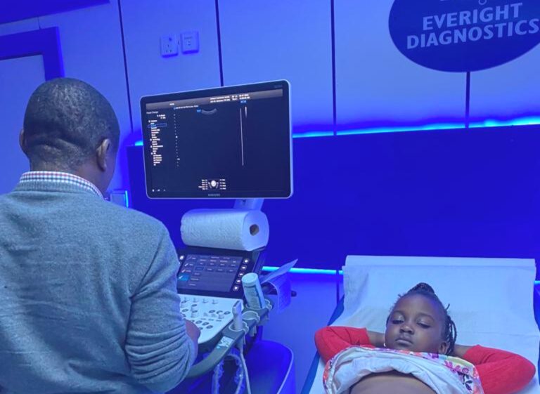
Pediatric echocardiography is ultrasonography used to evaluate the anatomical structure and function of the heart in children. An echocardiogram is a test that uses sound waves (ultrasound) to create images of the heart. Echocardiography is the most common test used in children to diagnose or rule out heart disease and also to follow children who have already been diagnosed with a heart problem. This test can be performed on children of all ages and sizes including foetuses and newborns. Echocardiography diagnoses cardiac problems and also guides heart surgery and complex cardiac catheterizations. Several cardiac conditions may be detected by echocardiography, including the following:
- Heart murmur
- Cardiomyopathy
- Congenital heart disease
- Congestive heart failure
- Pericarditis
- Valve disease
The ultrasound test is done with your child lying down comfortably on a bed. Infants may be able to lie in their parent’s lap. A small amount of clear gel is used between the transducer and chest to make sure there is proper contact. The gel doesn’t stain and is wiped off when the test is over. You’ll be able to see and hear the beating heart, as well as the flow of blood. Echocardiographic examinations take 20 to 40 minutes. It is important for the child to remain still to obtain an examination of acceptable quality. Younger children may require sedation to allow the cardiologist to obtain a complete study. If sedation is needed, the child will be asked not to eat or drink for several hours prior to testing. Older children and teenagers usually don’t need sedation and may enjoy watching the test. Except for allowing for the possibility of sedation, you don’t need to make any special preparations.
- Digital Radiography (X-ray)
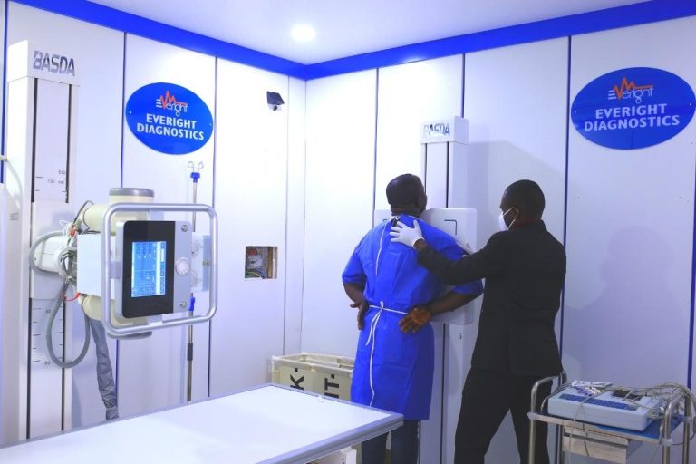
X-ray is the conventional and oldest medical imaging tool. They are used to view images of the bones and other structures in the body. X-rays can detect fractures, infections (pneumonia, osteomyelitis), calcifications (kidney stones), tumours, arthritis, and bone loss. Our digital x-ray system provides high picture quality and ensures accurate diagnosis.
- CT Scan
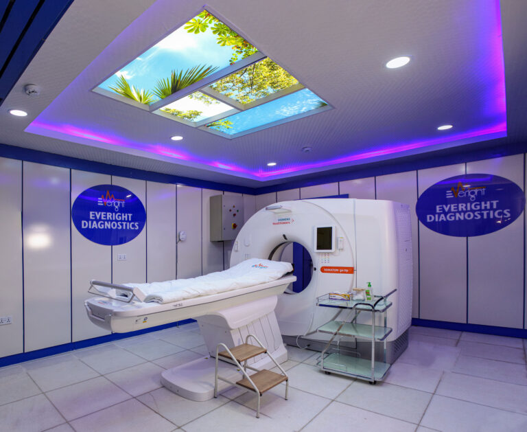
A CT scanner is like an advanced x-ray machine. It produces better images that provide more detailed information than plain X-rays. We have a 64 SLICE CT Scanner, which uses a series of X-ray images taken from different angles around the body and computer processing to create cross-sectional images (slices) of the bones, blood vessels and soft tissues. It is a vital medical diagnostic tool when fast scans are needed. A 64-slice machine can scan the whole body in 60 seconds!
- High-definition image quality with less dose and efficient workflow
- Trusted performance with high uptime
- MRI
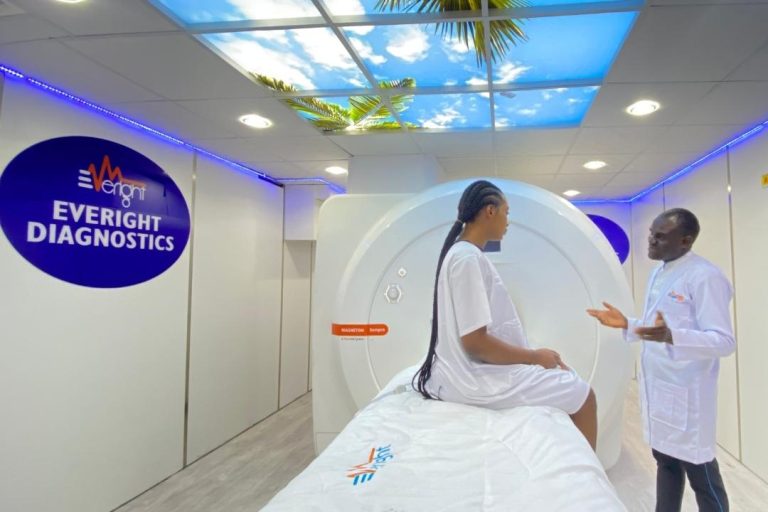
Alongside X-ray and CT scan images, we also have Magnetic Resonance Imaging (MRI). MRI is a large tube that contains powerful magnets. It uses strong magnetic fields and radio waves to produce a detailed image of organs and structures inside the body. These images help in diagnosing a variety of conditions, from torn ligaments to tumours. It is safe, as it does not use ionising radiation; a baby in the womb can get an MRI scan. It can be used for any part of the body but is excellent for the soft parts, such as the brain, muscles, ligaments, tendons, internal organs and blood vessels. A 1.5 Tesla brand of MRI makes the scan time as short as possible, gives excellent picture quality, thereby ensuring accurate diagnosis.
In our facility, we use an MRI scanner for:
- Brain and spinal cord anomalies
- Breast cancer screening
- Irregularities like tumours, cysts in various parts of the body
- Liver and other abdominal organs diseases
- Injuries to the joints in the body
- Heart and Vascular problems
- Pelvic pain evaluation in women
- Prostate evaluation
- Age determination
- Digital Mammography
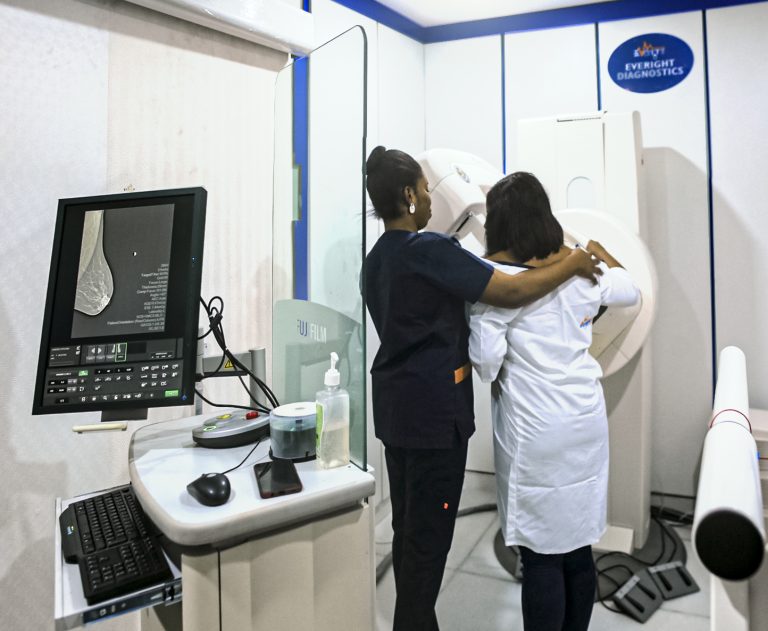
Physicians sometimes prefer mammography in breast cancer diagnosis, especially in women older than 40 years. Mammography uses low-dose X-rays to produce breast images known as mammograms. It is a screening tool for possible breast diseases.
- Electroencephalography (EEG) and Electrocardiogram (ECG)
We also have facilities for EEG and ECG.
EEG is a monitoring method used to record the electrical activity of the brain. This helps in determining changes in brain activity that might be useful in diagnosing and treating brain disorders and diseases (epilepsy).
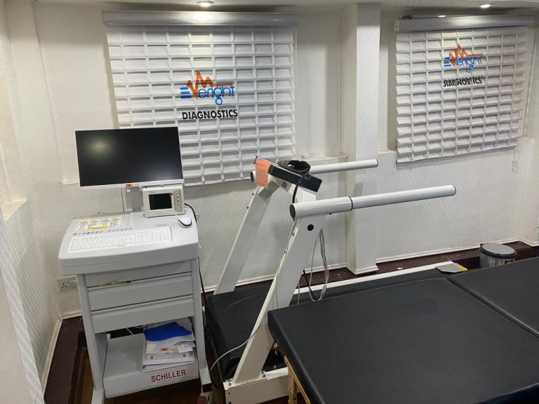
Electrocardiogram (ECG/EKG) is a medical diagnostic tool that measures the electrical activities of the heart. ECG is recommended for all adults, especially those with a family history of hypertension.